


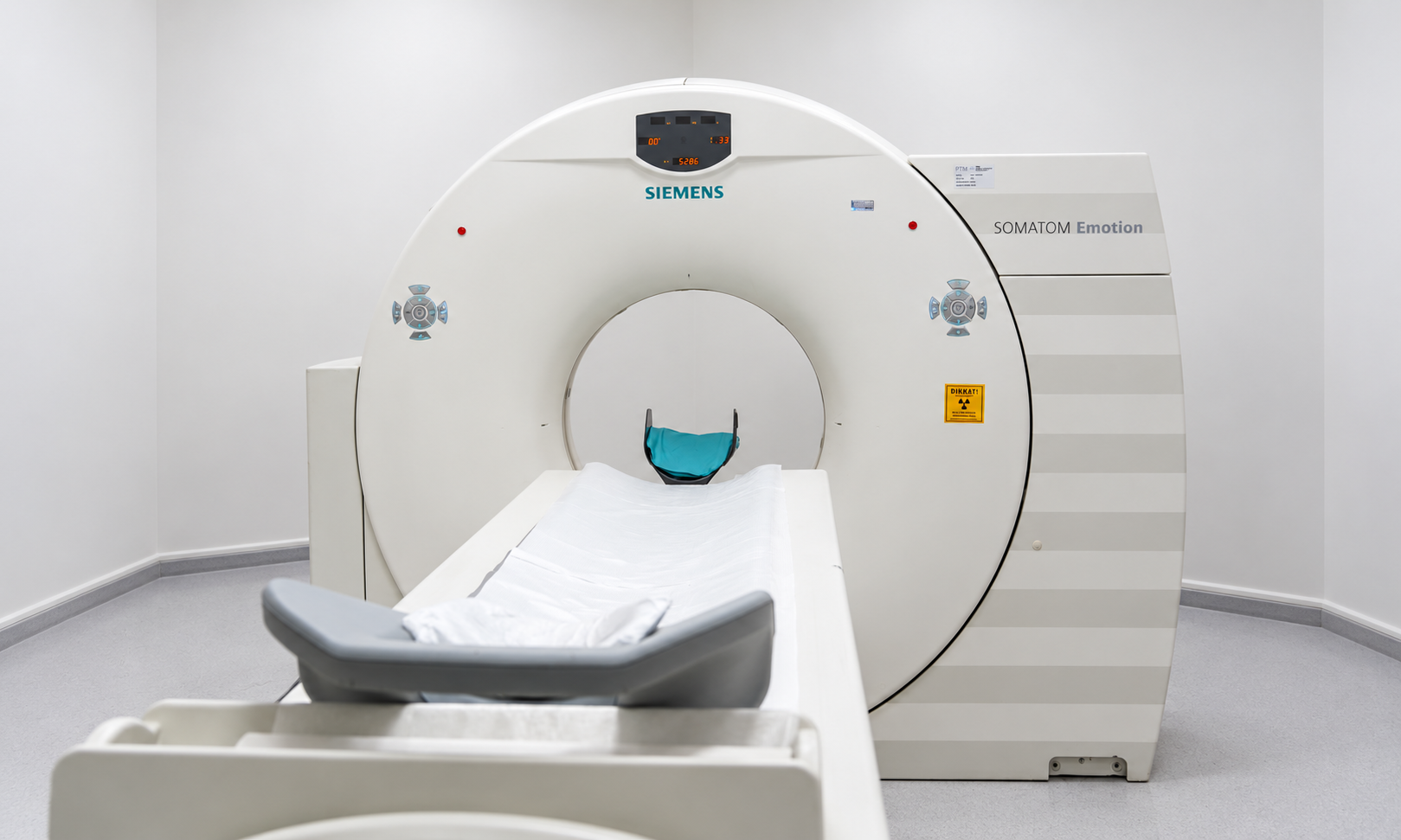
Radiology and Imaging supports diagnosis and follow-up by visualizing internal body structures using different technologies. Ultrasound, X-ray, computed tomography (CT), magnetic resonance imaging (MRI) are selected according to the clinical question. No single test is appropriate for every patient; the expected diagnostic benefit is considered together with potential issues such as ionizing radiation, contrast agents and individual safety factors.
What Is Radiology and Imaging?
Imaging does not always establish a diagnosis by itself. Findings are interpreted together with symptoms, physical examination and laboratory results. Different methods may be used for the same anatomical region because each technique provides different strengths for particular tissues and conditions.
For tests that use ionizing radiation, it is important to avoid unnecessary repetition, compare previous studies and report a possible pregnancy before imaging. MRI does not use ionizing radiation, but its strong magnetic field means certain metallic implants and medical devices require specific safety assessment.
What Symptoms or Clinical Questions May Lead to Imaging?
- Pain, swelling or trauma for which a physician requests imaging
- Need for abdominal, thyroid, breast or soft-tissue ultrasound
- X-ray assessment of lungs or bones
- MRI assessment of the brain, spine, joints or soft tissues
- CT for urgent or detailed cross-sectional imaging
- Breast screening or diagnostic imaging
- Doppler ultrasound assessment of blood vessels
Which Conditions May Be Evaluated?
- Musculoskeletal conditions: Imaging of fractures, joints, muscles, tendons and the spine.
- Abdominal and pelvic organ conditions: Assessment of the liver, gallbladder, kidneys, uterus, ovaries and other organs.
- Brain and nervous-system conditions: Structural evaluation with CT and MRI.
- Lung and chest conditions: Appropriate imaging, including X-ray and CT.
- Breast conditions: Ultrasound and, when indicated, MRI.
- Vascular conditions: Doppler ultrasound and appropriate CT/MR angiographic techniques.
How Does the Imaging Process Work?
Imaging is selected according to the referring clinician’s question and the patient’s individual circumstances. Not every modality is appropriate or necessary for every patient.
- Ultrasonography and Doppler ultrasonography
- Plain X-ray
- Computed tomography (CT)
- Magnetic resonance imaging (MRI)
- Contrast-enhanced imaging when clinically indicated
Service Areas
The following items describe common radiology services.
- Planning an imaging protocol appropriate to the physician’s request and clinical question
- Interpretation and reporting by a radiologist
- Comparison with previous examinations when images are available
- Medical-suitability assessment when contrast material is required
When Should You Seek Emergency Care Instead?
An imaging department is not a substitute for emergency treatment. With sudden stroke symptoms, major trauma, severe chest pain or severe abdominal pain, seek emergency medical care directly. The emergency team will determine which imaging test, if any, is required after the initial assessment.
Frequently Asked Questions
Are MRI and CT the same thing?
No. CT uses X-rays, whereas MRI creates images using a strong magnetic field and radiofrequency signals. The most appropriate method depends on the body region and clinical question.
Does ultrasound use radiation?
No. Diagnostic ultrasound uses high-frequency sound waves and does not use ionizing radiation.
Can X-ray or CT be performed during pregnancy?
When imaging uses ionizing radiation, the need for the test and possible alternatives are considered carefully during pregnancy. Pregnancy or possible pregnancy should always be reported to the healthcare team before the examination.
Why must metal objects be removed before MRI?
MRI uses a strong magnetic field. Some metal objects can create a safety risk or degrade image quality, and implants or medical devices may require specific compatibility checks before scanning.
What is contrast material?
Contrast agents can make certain tissues and blood vessels more visible on imaging. Kidney function, allergy history and the type of contrast agent are among the factors that may be reviewed before administration.
What does a radiology report mean?
The report describes findings identified by the radiologist and may include an interpretation or differential considerations when appropriate. The result should be assessed by the referring physician together with the clinical examination and other tests.
Should I bring previous scans?
Yes, if available. Previous images and reports can be valuable for comparison and may help determine whether a finding is new, longstanding or changing over time.
Does every pain require an MRI?
No. Imaging decisions depend on the examination and clinical suspicion. Some problems need no imaging, while in other cases X-ray, ultrasound or CT may be more appropriate than MRI.